
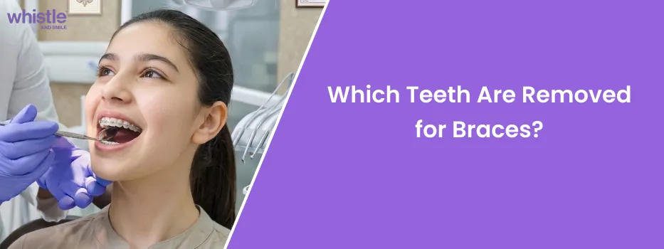
Which Teeth Are Removed for Braces? Orthodontic Extraction Protocols and Facts
 Medically Reviewed by Whistle Dental Team
When tooth extraction is required for orthodontic alignment, the teeth most commonly removed for braces are the first premolars (bicuspids), located directly behind the canines in each quadrant of the dental arch.
In specific clinical scenarios, orthodontists may alternatively extract second premolars, wisdom teeth (third molars), a single lower incisor, or structurally compromised teeth with extensive decay or trauma.
Key Takeaways
- The Primary Extraction Candidates: First premolars (bicuspids, teeth #4 and #5) are the most frequently extracted teeth because their central arch position creates ideal space to resolve crowding and retract flared incisors.
- Alternative Extraction Strategies: Second premolars are selected when less anterior retraction is desired to preserve facial fullness, while lower incisors or wisdom teeth are targeted for specific bite or space discrepancies.
- Why Canines Are Never Removed: Canine teeth (eye teeth) serve as the vital cornerstones of the dental arch, supporting the facial lip corners and providing essential canine guidance during chewing; they are preserved whenever possible.
- Predictable Gap Closure Timeline: Extraction spaces do not leave permanent gaps; orthodontic braces or clear aligners begin closing spaces within 2 to 3 months, achieving complete space closure within 6 to 12 months.
- In-Person Orthodontic Anchorage Control: Closing extraction spaces requires precise bodily translation and root torque management, necessitating direct clinical supervision by an MDS Orthodontist rather than direct-to-consumer mail kits.
Symmetrically positioned midway between front and back teeth, removing premolars generates immediate space to relieve severe crowding, retract protruding incisors, and correct bite discrepancies without compromising facial profile aesthetics under specialist orthodontist care.
Which Teeth Are Removed for Braces? The Orthodontic Selection Hierarchy
Orthodontic treatment aims to create a harmonious, functional bite and an aesthetically balanced smile. When there is a severe mismatch between tooth size and jaw size (arch length discrepancy), non-extraction methods like expansion reach biological bone limits. In such cases, orthodontists must select specific teeth for extraction based on a strict, evidence-based clinical hierarchy:
1. First Premolars (Bicuspids, Teeth #14, #24, #34, #44)
First premolars are the gold-standard choice in orthodontic extraction therapy. Located fourth from the front midline, directly behind the canines, these teeth sit precisely midway along the dental arch. Removing first premolars provides immediate, substantial space (approximately 7 to 8 millimeters per side) adjacent to crowded or protruding front incisors, allowing orthodontists to unravel overlapping teeth and retract flared crowns with maximum biomechanical efficiency.
2. Second Premolars (Teeth #15, #25, #35, #45)
Second premolars, positioned just behind the first premolars, are selected when a patient requires moderate space to resolve crowding but already has a relatively flat or balanced facial profile. By removing the second premolar instead of the first, more posterior molar anchorage is consumed as back teeth drift forward, preventing excessive retraction of the front teeth and protecting youthful lip fullness.
3. Wisdom Teeth (Third Molars, Teeth #18, #28, #38, #48)
Wisdom teeth are rarely extracted to create direct space for crowded front teeth during active treatment. Instead, third molars are removed when they are impacted, infected (pericoronitis), or physically obstructing the distalization (backward movement) of the entire molar dentition in non-extraction clear aligner protocols.
4. Single Lower Incisor (Tooth #31 or #41)
In select adult patients with severe lower anterior crowding, mild Class III tendency, or a Bolton tooth-size discrepancy (where lower teeth are disproportionately large compared to upper teeth), an orthodontist may remove a single lower central incisor. This conservative option resolves lower crowding rapidly without disturbing the stable posterior molar bite.
5. Severely Damaged, Decayed, or Compromised Teeth
Whenever possible, orthodontists avoid extracting pristine, healthy virgin teeth. If a patient possesses a first permanent molar or premolar that is heavily decayed, fractured, structurally weakened by endodontic therapy, or affected by severe localized periodontal bone loss, that compromised tooth is prioritized for extraction instead, preserving the patient's healthy teeth.
6. Over-Retained Baby Teeth and Supernumerary (Extra) Teeth
In children and teens, a primary milk tooth that fails to resorb its roots blocks the permanent tooth beneath it, forcing it to erupt ectopically. Similarly, supernumerary teeth (such as a midline mesiodens) introduce physical obstructions that crowd the arch. Removing these ectopic units clears the pathway for natural alignment.
Why Are Canine Teeth Never Extracted for Braces?
Common Clinical Extraction Patterns in Orthodontics
Depending on the diagnostic classification of malocclusion, orthodontists configure specific extraction patterns:
- Symmetrical Four-Premolar Extraction (All First Premolars): The classic protocol for severe bimaxillary protrusion or extreme generalized dental crowding. Removing four first premolars balances both upper and lower arches symmetrically, allowing front teeth to retract evenly and lips to close effortlessly.
- Two Upper First Premolars Only: Indicated in Class II Division 1 malocclusions (severe overjet) where the lower jaw is well-aligned and normal in size, but the upper front teeth project significantly forward. Removing only upper premolars allows the upper incisors to retract into a harmonious relationship with the lower teeth without altering the lower arch.
- Four Second Premolars: Selected when mild-to-moderate crowding is present and the facial profile is already straight or concave. This pattern allows back molars to move forward while minimizing backward retraction of the anterior smile line.
- Asymmetrical Extraction: Used in cases of severe dental midline deviation or unilateral crossbites, where teeth are extracted on only one side or in opposite quadrants to restore facial and dental symmetry.
Teeth Selected for Orthodontic Extraction
| Tooth Type | Arch Position | Primary Clinical Purpose | Effect on Facial Profile | Extraction Frequency |
| First Premolars | Between canines and 2nd premolars | Maximum space to unravel severe crowding and retract flared incisors | Permits controlled lip retraction, reducing profile convexity | Most common (80-90% of extraction cases) |
| Second Premolars | Between 1st premolars and molars | Moderate space creation; allows molars to drift forward | Preserves lip fullness; prevents sunken profile in flat faces | Common in moderate crowding |
| Wisdom Teeth | Terminal ends of both dental arches | Relieves posterior impactions; enables whole-arch molar distalization | Zero effect on facial structure or cheek fullness | Common adjunctive procedure |
| Lower Incisor | Lower front anterior center | Resolves severe localized mandibular crowding or Bolton discrepancy | Minimal to zero effect on outer profile | Select adult cases only |
| Compromised Molars | Posterior chewing zone | Removes severely decayed or root-fractured units instead of healthy teeth | Maintains natural tooth health without extra trauma | Case-specific strategic choice |
How Are Extraction Gaps Closed? The Biomechanical Process
A primary concern among patients is whether tooth extraction leaves permanent empty gaps in their smile. In orthodontic treatment, extraction gaps are deliberately and completely closed:
Orthodontists utilize two primary biomechanical methods to close extraction spaces: sliding mechanics and loop mechanics. In sliding mechanics, an orthodontic archwire guides teeth backward using elastomeric power chains or nickel-titanium (NiTi) closing coil springs. In loop mechanics, specialized wire loops gradually flex and close as teeth translate through bone.
To prevent unwanted movement of back molars (loss of anchorage), orthodontists often place Temporary Anchorage Devices (TADs). These miniature biocompatible titanium pins provide fixed, immovable bone anchors, allowing front teeth to retract fully without pulling back teeth forward.
Modern clear aligner systems (such as Whistle Clear Aligners) are also fully capable of closing extraction spaces. Through digitally staged movements and composite attachments placed on adjacent teeth, clear aligners pull crowns and roots together in a controlled, upright orientation without unwanted tipping.
How Long Do Extraction Gaps Take to Close?
- First 1 to 2 Weeks: The extraction socket forms a stable blood clot, and mucosal gum tissue rapidly epithelializes over the bone socket. Braces brackets are bonded or clear aligner trays begin active wear.
- Months 1 to 3: Initial orthodontic leveling and alignment occur. Flared incisors begin de-rotating, and early space closure becomes visible.
- Months 6 to 12: Active space closure mechanics translate teeth bodily across the extraction site. For most patients, visible spaces are completely closed within 6 to 12 months, well before the final detailing phase of treatment begins.
Non-Extraction Alternatives: When Can Removal Be Avoided?
Whenever clinically viable, orthodontists prefer non-extraction protocols. In mild to moderate crowding cases, alternative space-creating techniques include:
- Interproximal Reduction (IPR): Gently polishing 0.1 to 0.3 mm of enamel between contact points creates 2 to 4 mm of total space across the arch, resolving mild crowding without extractions.
- Arch Expansion: Widening narrow dental arches uprights inward-leaning teeth and creates arch perimeter space in patients with adequate alveolar bone thickness.
- Molar Distalization with Clear Aligners: Moving molars backward one by one using sequential clear aligners and mini-screws (TADs) frees up anterior space, eliminating the need for premolar removal in border-line cases.
Critical Safety Precautions and Anti-DIY Warnings
Tooth extraction and orthodontic space closure are delicate biological procedures that require licensed specialist oversight:
- Never Attempt Home Extractions: Attempting to pull or loosen teeth at home with tools or string introduces severe bacterial infections, fractures delicate jawbone plates, and permanently severs nerve supplies.
- Ban DIY Gap Bands and Elastics: Placing household rubber bands around teeth to close gaps causes the bands to creep beneath the gumline, destroying the periodontal ligament and causing catastrophic tooth loss within weeks.
- Avoid Unmonitored Mail-Order Aligner Kits: Direct-to-consumer mail-in aligner companies lack the clinical capability to close premolar extraction spaces safely. Moving teeth across large gaps without in-person root torque control causes severe root tipping, traumatic occlusal collisions, and permanent bone defects.
Your Next Steps
Determining which teeth are removed for braces is an evidence-based clinical decision that prioritizes long-term oral health, stable occlusion, and balanced facial aesthetics. First premolars remain the primary choice for relieving severe crowding and retracting protrusion, while second premolars, wisdom teeth, and lower incisors serve specialized clinical roles.
With modern orthodontic mechanics, extraction gaps close completely within 6 to 12 months, and computer-guided planning ensures your facial profile remains youthful and natural. By partnering with a qualified orthodontist, you can achieve a radiant, functionally sound smile that lasts a lifetime.
Book 3D Teeth Scan
Consult our experts and get a 3D preview of your smile transformation!
Which Teeth Are Removed for Braces
FAQ's
Q1.Which specific teeth are most commonly pulled for braces?
First premolars (bicuspids, located directly behind the canines) are by far the most commonly extracted teeth for braces because their central position in the arch creates ideal space to relieve crowding and retract flared front teeth.
Q2.Is it better to remove 2 teeth or 4 teeth for braces?
Neither option is inherently better; the choice depends on your specific bite and crowding. Symmetrical four-premolar extraction is standard for bimaxillary crowding or protrusion, while two upper premolars are removed to camouflage severe overjets where the lower arch is already well-aligned.
Q3.Do braces go on before or after teeth are removed?
Both clinical sequences are common. Some orthodontists place braces first to level the teeth before extracting, while others extract premolars 1 to 2 weeks prior to bonding brackets to utilize the immediate space from day one. Your orthodontist will tailor the sequence to your movement plan.
Q4.Can clear aligners close extraction spaces as effectively as braces?
Yes. Whistle Clear Aligners utilize custom digital staging and composite attachments to translate teeth bodily across extraction spaces without tipping, provided patients wear trays for 20 to 22 hours daily under doctor supervision.
Q5.Can I avoid tooth extraction by expanding my jaw instead?
Arch expansion can resolve mild to moderate crowding, but it cannot fix severe arch length discrepancies or heavy protrusion without pushing tooth roots outside the supporting alveolar bone. Your orthodontist will evaluate your 3D scan to determine safe biological boundaries.
You May Also Like


 Medically Reviewed by Whistle Dental Team



















 Medically Reviewed by Whistle Dental Team

 Medically Reviewed by Whistle Dental Team