

White Spots on Teeth: Causes, Treatment Options and Prevention
 Medically Reviewed by Whistle Dental Team
White spots on teeth are chalky patches caused by enamel demineralization, fluorosis, or developmental defects, and can be treated through remineralization, resin infiltration, microabrasion, or cosmetic dental procedures.
Clinically presenting as incipient white spot lesions, dental fluorosis, or enamel hypoplasia, these patches commonly emerge from trapped bacterial plaque around braces, excess childhood fluoride ingestion, or chronic acidic diets.
Key Takeaways
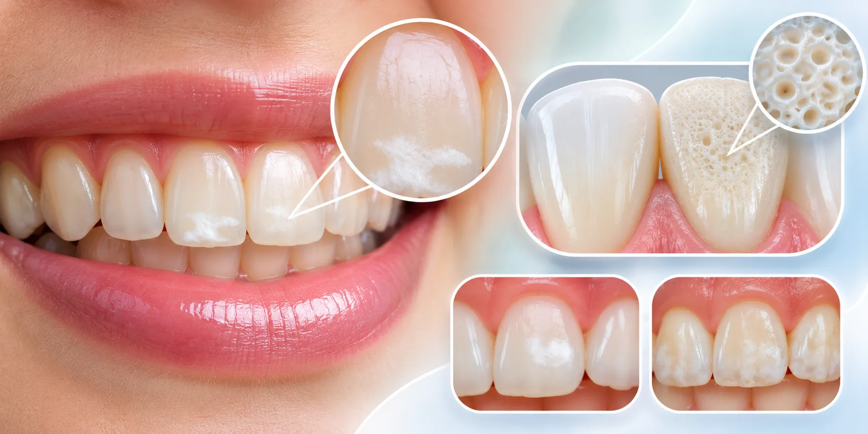
- Subsurface Enamel Porosity: White spots develop when enamel loses calcium and phosphate crystals, creating microscopic pores that scatter light and appear opaque or chalky.
- Root Cause Dictates Treatment: Incipient white spot lesions from plaque demineralization can be remineralized, whereas developmental fluorosis or hypoplasia require microabrasion, resin infiltration, or aesthetic bonding.
- Drill-Free Resin Infiltration: The ICON resin infiltration technique eliminates white lesions in a single visit without dental drilling by infusing optical-grade liquid resin into porous enamel.
- The Teeth Whitening Paradox: Over-the-counter bleaching cannot remove white spots and frequently increases color contrast initially, making spots stand out before surrounding enamel blends.
- Orthodontic Shielding: Fixed metal braces create difficult plaque traps that cause bracket-shaped white outlines; removable clear aligners allow unhindered daily brushing and flossing to keep enamel intact.
Depending on lesion depth, effective clinical pathways on how to get rid of white spots on teeth range from non-invasive remineralization therapy and micro-invasive resin infiltration (ICON) to enamel microabrasion, cosmetic bonding, and lifestyle adjustments that permanently protect your smile.
What Are White Spots on Teeth?
Primary Causes of White Spots on Teeth
- Plaque Accumulation and Enamel Demineralization: Bacterial biofilm metabolizes dietary sugars and releases lactic acid, dropping oral pH below the critical threshold of 5.5. This acidic environment leaches calcium and phosphate ions from subsurface enamel, creating an incipient white spot lesion: the earliest reversible stage of a dental cavity.
- Orthodontic Complications (Post-Braces White Scars): Fixed brackets, archwires, and elastomeric bands create complex stagnation areas where food debris accumulates. When meticulous sulcular brushing is hindered, demineralization occurs along bracket perimeters, leaving permanent square or rectangular white outlines once braces are debonded.
- Dental Fluorosis: Ingesting excessive systemic fluoride during early childhood (specifically between birth and age eight while amelogenesis is active) disrupts ameloblast protein processing. This results in subsurface enamel hypomineralization, typically presenting as symmetrical lacy white lines, flecks, or chalky horizontal bands across contralateral teeth.
- Enamel Hypoplasia and Molar Incisor Hypomineralization (MIH): Systemic stressors during early childhood, such as severe maternal nutritional deficits, high fevers, infectious illnesses, premature birth, or direct trauma to primary incisors, disrupt enamel matrix secretion. Erupted permanent teeth exhibit demarcated white, yellow, or brown opacities with compromised structural hardness.
- Temporary Enamel Dehydration: Mouth breathing during sleep or professional teeth whitening temporarily removes moisture from superficial enamel micropores. This transient dehydration alters light refraction, creating harmless white patches that completely fade within a few hours as saliva rehydrates the enamel.
How to Get Rid of White Spots on Teeth: Clinical Treatment Pathways
Modern restorative dentistry offers multiple conservative and non-invasive methods to eliminate, blend, or conceal enamel opacities based on their depth and origin:
1. Remineralization Therapy (Non-Invasive)
For early active white spot lesions where the outer enamel surface remains intact, non-invasive remineralization is the primary clinical line of defense. Applying professional 5% sodium fluoride varnishes (22,600 ppm), high-concentration prescription toothpastes (5,000 ppm NaF), and Casein Phosphopeptide-Amorphous Calcium Phosphate (CPP-ACP / MI Paste) floods the enamel with bioavailable calcium and phosphate ions, reforming robust fluorapatite crystals that halt decay progression and diminish chalkiness.
2. Micro-Invasive Resin Infiltration (ICON Technique)
Resin infiltration represents a revolutionary bridge between prevention and restoration, resolving white spot lesions and mild fluorosis in a single painless appointment without any drilling or anesthesia. The clinician conditions the hyper-mineralized surface layer with a mild 15% hydrochloric acid gel, dries the porous lesion with ethanol, and infuses a low-viscosity optical resin directly into the subsurface micro-porosities.
Because the cured resin has the exact same light refractive index as healthy natural enamel, the white spot visually disappears, instantly blending into the natural tooth shade.
3. Enamel Microabrasion
Ideal for superficial fluorosis and shallow extrinsic developmental defects, enamel microabrasion combines a mild hydrochloric acid solution with fine pumice or silicon carbide particles. The clinician gently abrades the outer 100 to 200 micrometers of stained enamel using a slow-speed rubber cup. This controlled mechanical-chemical erosion eliminates surface discolorations and compacts the outer enamel layer, leaving a lustrous, smooth finish.
4. Controlled Professional Whitening (Bleaching)
While professional whitening does not chemically dissolve white spots, it safely lightens the surrounding natural tooth structure to match the blemish, dramatically reducing contrast. Patients must be aware of the temporary whitening paradox: whitening gels initially dehydrate enamel, making white spots appear brighter for the first 24 to 48 hours before natural rehydration and uniform shade stabilization occur.
5. Cosmetic Composite Bonding and Porcelain Veneers
For deep, extensive enamel hypoplasia or structurally compromised teeth where microabrasion or resin infiltration cannot achieve optical harmony, aesthetic restorative care is indicated. Direct composite bonding involves placing tooth-colored resin over the lesion, while custom laboratory-fabricated ultra-thin porcelain veneers provide a durable, lifelike aesthetic shield across the entire facial surface.
White Spot Types and Clinical Solutions
| Condition Type | Underlying Etiology | Visual Appearance | Enamel Integrity | Recommended Clinical Pathway |
| Incipient White Spot Lesion | Bacterial plaque acids, post-braces demineralization | Chalky white, dull, along gumline or bracket margins | Demineralized subsurface, porous outer surface | Fluoride varnish, CPP-ACP remineralization, ICON resin infiltration |
| Dental Fluorosis | Excessive systemic fluoride intake during tooth formation | Bilateral, symmetrical lacy white flecks, horizontal streaks | Hypomineralized subsurface, intact hard exterior | Enamel microabrasion, ICON resin infiltration, professional whitening |
| Enamel Hypoplasia | Childhood high fever, nutritional deficiency, early trauma | Demarcated opaque white, cream, or brown pits/grooves | Structurally thin, brittle, prone to rapid breakdown | Direct composite bonding, aesthetic porcelain veneers |
| Enamel Dehydration | Mouth breathing during sleep, recent bleaching gels | Uniform, faint chalky patches across incisal edges | Structurally normal, transient water loss | Natural salivary rehydration, neutral fluoride hydration rinse |
Orthodontic White Spot Lesions: Braces vs Clear Aligners
The clinical incidence of white spot lesions during traditional fixed orthodontic treatment ranges between 30 and 70 percent. Fixed brackets and wires physically obstruct normal mechanical toothbrushing, while creating countless micro-crevices where plaque biofilm stagnates. If hygiene falters, localized enamel demineralization leaves permanent chalky outlines around each bracket.
In contrast, modern clear aligner therapy dramatically lowers the risk of white spot formation. Because Whistle Clear Aligners are completely removable, patients can brush and floss their natural teeth with 100 percent access using standard sulcular techniques.
However, one essential discipline is mandatory: patients must thoroughly brush their teeth before snapping aligners back in. Putting aligners over dirty teeth seals trapped fermentable carbohydrates and acidic bacteria directly against enamel for hours, accelerating white spot formation.
Proven Prevention Strategies for Lifelong Enamel Protection
Preventing new white spots from developing requires consistent preventive hygiene habits and proactive dietary choices:
- Sulcular Brushing Twice Daily: Brush for two full minutes twice daily using a soft-bristled brush angled at 45 degrees toward the gumline, using fluoride toothpaste containing 1000 to 1450 ppm fluoride.
- Daily Interdental Biofilm Control: Clean between teeth every night with dental floss, interdental proxy brushes, or an orthodontic water flosser, removing plaque from contact zones where toothbrushes cannot reach.
- Regulate Dietary Acids and Sugars: Minimize constant snacking and sipping on carbonated soft drinks, packaged fruit juices, and sports drinks. Continuous sipping maintains an acidic oral pH, keeping enamel under constant acid attack.
- Post-Acid Water Rinsing Protocol: After consuming acidic food or beverages, rinse vigorously with plain water immediately. Wait at least 30 minutes before brushing with toothpaste to prevent mechanically abrading softened enamel.
- Pediatric Fluoride Guidelines: For children under age three, use a tiny smear of fluoride toothpaste (rice-grain sized). For children aged three to six, use a pea-sized amount and supervise spitting to prevent systemic ingestion.
- Biannual Professional Prophylaxis: Schedule dental checkups and professional ultrasonic cleaning every six months to eliminate calcified plaque (calculus) and apply topical remineralization varnish.
When to Consult a Dentist or Specialist
Self-diagnosis cannot accurately determine whether an enamel blemish is an active carious lesion, systemic fluorosis, or structural hypoplasia. Seek prompt professional evaluation if you notice:
- Rapidly Spreading Opacities: White spots that expand or multiply within weeks indicate active, aggressive acid demineralization requiring immediate remineralization.
- Color Shift to Yellow or Brown: When a white spot transitions to brown or dark grey, it signals advanced bacterial penetration and active cavitation.
- Surface Roughness or Cavitation: If you feel a noticeable pit, groove, or catch with your fingernail or tongue, enamel collapse has occurred and restorative intervention is needed.
- Thermal or Sweet Sensitivity: Pain or sharp twinges when consuming cold drinks or sweet snacks indicate that mineral loss has compromised the enamel barrier and affected the underlying dentin.
In-Person Clinical Supervision Across Whistle Partner Clinics
Safely diagnosing and treating white spot lesions requires advanced clinical instrumentation, including intraoral magnification, transillumination diagnostics, and specialized remineralizing chemistry. Whistle operates in partnership with Clove Dental across 600+ physical partner clinics nationwide, providing patients with access to verified dental surgeons and MDS Orthodontists.
Whether evaluating post-orthodontic enamel demineralization, performing gentle resin infiltration, or planning clear aligner therapy that protects your enamel, Whistle partner clinics combine cutting-edge dental technology with transparent payment plans and flexible monthly EMI options. Face-to-face clinical checkups ensure your journey to a balanced, radiant smile is scientifically sound and completely safe.
Ready for a Healthier, Brighter Smile?
White spots on teeth are common, highly treatable dental concerns that no longer require aggressive drilling or invasive crowns.
By understanding the critical distinction between active demineralization lesions, systemic fluorosis, and enamel hypoplasia, patients can pursue targeted, evidence-based treatments ranging from topical remineralization pastes and ICON resin infiltration to conservative microabrasion.
Most white spots can be improved once you know what's behind them. Book a 3D teeth scan at a Whistle partner clinic and have an expert take a look.
Also Read:
Book 3D Teeth Scan
Consult our experts and get a 3D preview of your smile transformation!
White Spots on Teeth
FAQ's
Q1.Can white spots on teeth go away on their own without treatment?
Transient dehydration spots caused by sleeping with an open mouth or recent whitening fade naturally within a few hours as saliva rehydrates enamel. However, true white spot lesions from plaque demineralization, fluorosis, or hypoplasia do not vanish spontaneously and require clinical remineralization or cosmetic resin infiltration.
Q2.Do white spots on teeth after braces ever fade?
Some post-orthodontic white lesions naturally soften and blend over several months as saliva and fluoridated toothpastes remineralize the surface. Stubborn, prominent lesions respond exceptionally well to micro-invasive resin infiltration (ICON) or enamel microabrasion.
Q3.Does professional teeth whitening get rid of white spots?
No. Professional whitening bleaches the natural surrounding enamel to a lighter shade, which visually minimizes the contrast so spots become less noticeable. However, whitening initially dehydrates enamel and may make spots appear brighter for the first 24 to 48 hours.
Q4.Why do children frequently develop white spots on their teeth?
In children, white spots usually stem from dental fluorosis (swallowing excess fluoridated toothpaste or drinking high-fluoride ground water before age eight) or enamel hypoplasia caused by early childhood illnesses and nutritional deficiencies.
Q5.Can baking soda or lemon juice remove white spots on teeth at home?
No. Applying lemon juice or scrubbing with baking soda severely damages enamel. Lemon juice contains citric acid that dissolves hydroxyapatite crystals, while baking soda is abrasive and wears away fragile enamel, worsening the blemish.

 Medically Reviewed by Whistle Dental Team



 Medically Reviewed by Whistle Dental Team







 Medically Reviewed by Whistle Dental Team